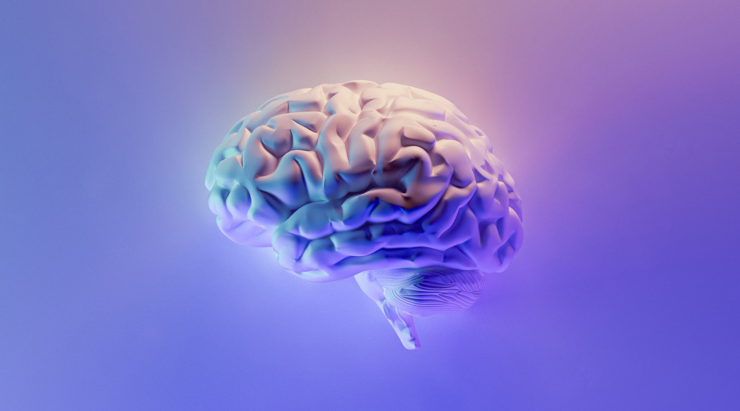
The brain is like an incredibly complex web of highways where information travels. These roads are composed of the brain cells – neurons and non-neuronal cells, including astrocytes and microglia – arranged in the structure formed by the non-cellular component known as the extracellular matrix.
The complexity of cellular arrangement and the connections made between them can lead to tortuous labyrinths hard to follow and understand, and when trying to study how the brain works, scientists have a hard time building models that closely mimic this intricate organization.
“The neurons in the brain are arranged in patterns, with regions rich in neuron cell bodies and other regions containing projections used to communicate with other neurons – often called white matter tracks,” said Helena Parkington, professor at the Department of Physiology at Monash University in Victoria, Australia. “The patterns facilitate optimal interconnection between the neurons to optimize communication and the formation of a 3D neural network.”
Trying to build a useful brain model of study, Parkington and a team of collaborators have turned to 3D bioprinting. In a recent study published in Advanced Healthcare Materials, they successfully employed bioprinting techniques to create 3D neuronal constructs using rat neurons and astrocytes.
“In this study we used extrusion 3D bioprinting, using a gelatin-PEG bioink, and constructed a brain structure similar to those that occur in the memory and learning region of the brain,” said Parkington. “We then tested the neurons and found that they performed in a manner similar to how they perform in the brain.” This innovative approach provides an exciting platform for investigating neural circuitry, engineering neuromorphic circuits, and conducting in vitro drug screening.
Printing a piece of brain
Bioprinting is a revolutionary technology that combines principles from 3D printing and tissue engineering to create living biological structures. It enables the precise deposition of living cells, biomaterials, and bioactive factors to fabricate functional tissues and organs.
In the case of neurons, the challenges lie in maintaining the viability and functionality of the printed cells that are highly delicate and require precise environmental conditions, such as appropriate nutrient supply and oxygenation. Moreover, ensuring proper cell-cell interactions and the formation of connections between neurons, called synapses, is also crucial for developing functional neural tissues.
To allow the proper interaction between cells, it is important to choose a good material that mimics the extracellular matrix where the cells grow, expand, and make synapses. One such class of materials are called “bioinks” and consist of a biocompatible hydrogel that provides structural support and nutrients for the cells. Parkington and the team used a bioink composed of gelatin-norbornene poly(ethylene glycol)-dithiol (or gelatin-PEG), which in a previous study they optimized to allow neuronal viability.
The team bioprinted rat neurons and astrocytes within the gelatin-PEG in eight layers, and allowed the growth of the cells for two weeks. When looking at the 3D printed morphology, they found that the neurons formed complex structures with other neurons and with the astrocytes and also expanded along the bioink.
“We found that the projections emanating from our neurons in the cellular layer readily travelled through the acellular layer [composed of gelatin-PEG] using it as a highway to communicate with neurons in other cellular layers,” said John Forsythe, professor at the Department of Materials Science and Engineering in Monash University, and co-lead author of the study. “A similar process is widespread in the normal human brain where there are cell rich layers connected by white matter tracks.”
A 3D printed model for brain studies
To assess the functionality of the printed neurons, the researchers conducted a series of analyses. The results revealed that the bioprinted neurons exhibited spontaneous electrical activity, a hallmark characteristic of healthy neural cells, indicating that the neurons were capable of generating electrical signals on their own, similar to neurons growing in a natural biological environment.
In addition, they observed that the neurons responded appropriately to various stimuli, validating their ability to process and react to external signals. These findings provided further evidence that the bioprinted neurons exhibited behavior akin to neurons growing in a native biological setting.
The successful maintenance of electrical activity in the bioprinted neurons represents a significant step forward in the field of neuroscience and bioprinting. It suggests that the printed neural tissue can recapitulate essential functions of natural neurons, thereby holding great promise for various applications.
“A 3D bioprinted model of the brain would have many benefits in drug and toxicity screening,” said Forsythe. “We are also interested in further studying neural network formation and building a living AI platform,” he added.
Reference: Yue Yao, et. al., 3D Functional Neuronal Networks in Free-Standing Bioprinted Hydrogel Constructs, Advanced Healthcare Materials (2023). DOI: 10.1002/adhm.202300801
Feature image by Milad Fakurian on Unsplash


