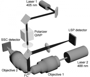
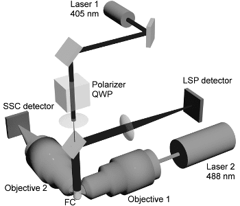
In a  new study, Valeri Malstev and coworkers from Novosibirsk, Russia, report a new method for simultaneous identification and characterization of microparticles (MPs), also known as extracellular vesicles (EVs) and platelets (PLTs) in the platelet-rich-plasma (PRP), improving MP characterization method and combining it with their previously developed method to measure PLTs volume and shape. First they improved the detection threshold of their scanning flow cytometer by replacing the 660 nm laser by the 405 nm and 488 nm lasers to measure the light-scattering profiles and side scattering from MPs, respectively. Finally they used two optical models, a sphere and a bisphere, to describe MPs shape and, hence, to differentiate single MPs from MP aggregates. The authors report that these modifications have significantly improved the accuracy of detection and characterization of MPs and PLTs in PRP, and also decreased the uncertainties of MP size and refractive index.
MPs are released from cellular membranes into blood and other body fluids under stress conditions. They are highly variable in size (100 nm to 1mM), cellular origin, function and other characteristics, and have been implicated in many biological properties and functions. MPs have been found in clinical samples for cardio-vascular diseases, cancer, sepsis, and auto-immune diseases, making them strong candidates as disease biomarkers, drug delivery vehicles and vaccines to diagnose and treat these diseases. Conventional FCM that uses light scattering has been used to study MPs for several decades, however, due to current technical limitations it cannot accurately detect and characterize MPs in sub-micrometer size range and phenotype MPs and PLTs. Therefore, the authors propose that their method is highly significant for biomedical research and diagnostics.


