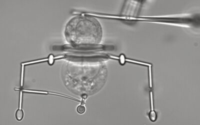
The inside of a cell is a busy place filled with a web of interactions between proteins, membranes, DNA, and other molecules to keep our bodies functioning.
Mapping these connections is vital for understanding health and how to repair this network when it is disturbed. Unfortunately, the tools to track proteins within a living cell struggle to keep up with the traffic.
To see proteins at work in real time, researchers are building a universal set of probes to find specific proteins among the organized chaos in order to explore some of the busiest corners of the cell.
Mapping proteins throughout a cell
Current protein visualization techniques are frozen in time and only work with dead and fixed cells, providing a single snapshot of protein interactions in a cell. Some live imaging techniques exist and rely on fluorescent antibodies designed to bind a protein and emit a colorful signal. However, the problem with antibodies is their size.
Within a cell there are sub-compartments enclosed in membranes called organelles. Proteins readily travel across these barriers and carry out important reactions within and between them, but because antibody probes are too large to breach these membranes, much of what goes on inside the organelles remains a mystery.
According to Xiaoding Lou, professor of materials science and chemistry at the China University of Geoscience, understanding the complete network of protein interactions inside a cell and its organelles is critical for understanding health and disease.
“Once the body undergoes pathological changes, the expression or activity of proteins will be affected,” she wrote in the paper published in Angewandte Chemie International Edition. “Therefore, realizing the protein analysis in the organelle of living cells is of great significance for developing diagnostic and therapeutic methods of major diseases.”
The new system consists of multiple molecules attached to a scaffold, each fulfilling a specific role: locating the organelles and proteins, penetrating the membrane, and producing a fluorescent signal.
The probe starts with a molecule called tetraphenylethylene or TPE and it is the scaffold onto which the functional molecules are added. These are the interchangeable protein and organelle recognition sites, fluorescent signaling molecules and a special membrane penetration component which, as the name suggests, helps the probe squeak through the intra-cellular membranes.
Probes that can get into organelles
This complex of molecules travels the cell and, depending on which recognition sites have been added, bind to proteins, and emit fluorescent light, like antibody probes. However, unlike antibodies, they can cross into the organelles and find their targets. Another improvement on antibodies is the ease with which recognitions sites can be changed.
“The universal detection platform can achieve protein analysis in different organelles through simple transformation of protein recognition and organelle targeting units,” wrote the scientists in their study. Meaning the scaffold remains the same and the units that find the organelles and proteins are interchangeable.
So far, recognitions units for six proteins and three unique organelle membrane molecules have proven effective in the lab. According to Lou, the probe could distinguish proteins throughout the cell and organelles, providing useful proteomic information. Furthermore, the ability to detect combinations of proteins in a cell can help discriminate between cell types, providing a quick and reliable cell fingerprint.
Now, more recognition sites can be designed, and the overall system optimized. “The signal-to-noise ratio needs to be further improved,” wrote the team. Along with recognition sites, the lab group is also exploring swapping out the fluorescent unit to emit different regions of the light spectrum which could improve the sensitivity and accuracy for certain proteins.
Reference: Xia Wu, et al., A Universal and Programmable Platform based on Fluorescent Peptide-Conjugated Probes for Detection of Proteins in Organelles of Living Cells, Angew. Chem. Int. Ed., (2024). DOI: 10.1002/anie.202400766
Feature image credit: National Cancer Institute on Unsplash

