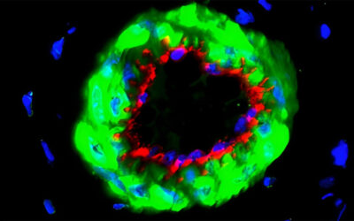
Impact of a retinal degenerative disease like macular degeneration and retinitis pigmentosa is debilitating due to partial or complete vision loss. The underlying reason is damage (age-related, genetic or environmental) to a thin layer of cells, the retinal pigment epithelium (RPE), located between the photoreceptor of the neural retina and the choroidal blood supply at the back of the eye. In an era where 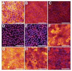 stem cell technology holds promise for robust disease-modeling and improved targeted cell-based therapies, effective treatment for a retinal degenerative disease is an unmet clinical need.
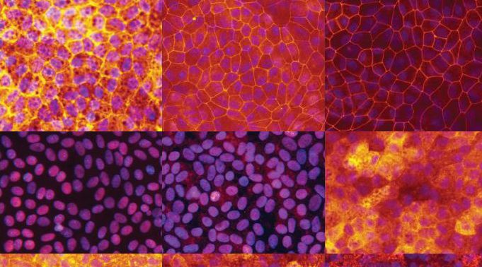
Addressing this problem, Fernandes and coworkers in the Blenkinsop laboratory at Icahn School of Medicine at Mount Sinai, New York, have developed a technique to create a human “RPE disease in a dish model”. In a recently published article in Current Protocols in Stem Cell Biology, they elaborate highly efficient and reproducible protocols for isolation and culture of adult human RPE stem cell-derived cells (ahRPE-SCs). In contrast to the traditional whole globes, this new protocol makes use of the posterior poles of globes used in corneal transplants for establishing RPE cultures with superior total cell yield and higher success rate.
The article includes a set of protocols for isolation, culture and expansion of ahRPE-SCs. The unit includes two protocols (immunofluorescence and transepithelial resistance measurements) to phenotypically characterize the established cell lines and assess whether the cultures mimic native RPE physiology. Adult human RPE stem cell-derived cells (ahRPE-SCs) are ideal for modeling retinal degenerative diseases and the use of these advanced protocols can provide a new source of high quality primary RPE line cultures.
To find out more please read: Fernandes, M., McArdle, B., Schiff, L., & Blenkinsop, T. A. (2018). Stem cell–derived retinal pigment epithelial layer model from adult human globes donated for corneal transplants. Current Protocols in Stem Cell Biology, 45, e53. doi: 10.1002/cpsc.53

