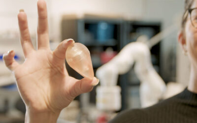
Researchers have adopted a specialized 3D bioprinting technique to create advanced models of the human brain, known as organoids. Their new approach retains the natural features of brain cell cultures, making it possible to achieve a higher level of organoid complexity than ever before.
Customizable, anatomically accurate brain organoids, like the one developed in this study, are potentially useful for many reasons, including studying neurological diseases and brain development in detail, as well as testing how drugs permeate and interact in the brain.
However, previously developed brain organoids have failed to realistically mimic the extracellular matrix, a crucial support structure made up of proteins and carbohydrates that provides a protective environment for the organoids to grow. This limits their usefulness.
“Brain organoid cultures are currently limited by a lacking ability to precisely control their extracellular environment,” the researchers stated in their paper. They also noted that the previous approaches are difficult to scale up.
Incomplete brain organoids, poor models
Without a realistic mimic of this matrix, the organoids may not develop or function in a way that truly represents the human brain.
Also missing from previous brain organoids is the brain’s network of blood vessels called the vasculature. But vascular cells, which account for about 10% of all cell types in the brain, are not produced from the same stem cells that generate other brain cells, like neurons. They need to be incorporated into organoids using a different approach.
“Most of the cell types within the brain are derived from a cell lineage that does notproduce vascular cells,” explained Steven Sloan, a lead researcher on the study and professor of human genetics at Emory University School of Medicine in Atlanta, Georgia.
“Therefore, unless these cells are physically integrated into the organoid, or genetic tricks are employed to force their generation, typical brain organoid cultures have no vasculature,” he elaborated.
Printing the organoid scaffolds
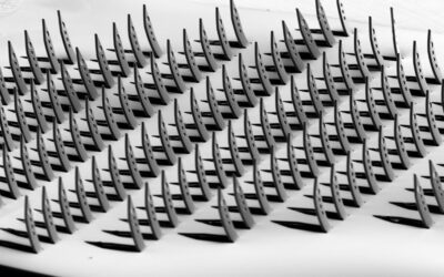
To precisely shape the extracellular matrix and effectively introduce vascular cells into brain organoids, Sloan and his team used a 3D printing technique known as embedded bioprinting.
In conventional 3D bioprinting, a “bioink” containing living cells and a biocompatible scaffold material is slowly forced through a nozzle at computer-programed pressures and speeds by the printhead — the part that contains the ink — in air, forming 3D objects.
But when cells are embedded in bioinks and printed directly, they may lose some of their innate abilities, like self-organization, which isn’t ideal. To avoid this possibility, Sloan and his team opted to print the scaffold first and then encapsulate the preformed organoid inside it.
The problem with direct printing is that many bioinks are not viscous enough to retain their shape, easily collapsing or deforming under gravity. To overcome this issue, the printhead can be immersed in a more viscous material — like gelatin, for example — as a support. This material can then be easily washed away after the printing process is complete.
“The high viscosity [of the support material] allows delicate structures (or intricate details like channels or pores) to maintain their structure throughout the printing process,” Sloan stated.
GelIMA scaffold provides a mimic
To print the organoid scaffold, the researchers chose a hydrogel as the viscous support material and included a gelatin-based material, known as GelMA, in the bioink formulation to mimic the extracellular matrix.
“GelMA is great because it’s biologically inert, cells are happy to grow on it, and it’s very easy to modify in the future by incorporating proteins or small molecules,” said Sloan.
They also programmed the printer to incorporate channels into the scaffold design, allowing them to load the organoid into the scaffold and separately introduce vascular cells.
They loaded the organoid, which they had grown beforehand from a culture of human-induced pluripotent stem cells, through the hydrogel’s top channel. After sealing the organoid in place using UV light, strengthening the scaffold, they coated the side channels with human umbilical vein endothelial cells. These stem cells gradually infiltrated the organoid in the center of the scaffold, vascularizing it.
To tune the composition and physical properties of the extracellular matrix-like scaffold, including its porosity and stiffness — which, in an extracellular matrix, vary from person to person and is affected by disease — the concentration of the gelatin-based material or UV irradiation time and intensity can be changed.
“These scaffolds can have almost any architecture desired and are reproducible at scale,” Sloan stated. “Most importantly, the organoids like being inside this bioprinted scaffold and develop normally.”
Where to now?
“While this is certainly not the first time that vascular cells have been ‘mixed’ with brain organoids, it’s a novel take on how to introduce vascularization that has the capacity to be functional,” Sloan said of their study.
According to Sloan, simply mixing vascular cells with an organoid doesn’t automatically translate into a functioning organoid. By incorporating channels into the scaffold and mimicking the flow of fluids into and out of the brain, their bioprinting approach allows for real functionality.
He and his team plan to update their model with this fluid-flow capability. “We don’t yet have a functional vascular system with a clear inlet and outlet [to create fluid flow into and out of the organoid], and that will need to be a major focus of next steps,” Sloan stated.
“Without that functional component, it does remain difficult to investigate important questions like blood–brain barrier formation, or even to think about screening drug permeability through brain vasculature,” he continued.
Sloan is also looking forward to adding more features to the model using their bioprinting approach.
“You can load all kinds of things in these bioinks, so there are many options in terms of integrating other signaling cues, growth factors, or even [long-range signaling molecules called] morphogens to developing organoids,” he shared with us.
Reference: Melissa A. Cadena et al. A 3D Bioprinted Cortical Organoid Platform for Modeling Human Brain Development. Advanced Healthcare Materials (2024). DOI: 10.1002/ adhm.202401603