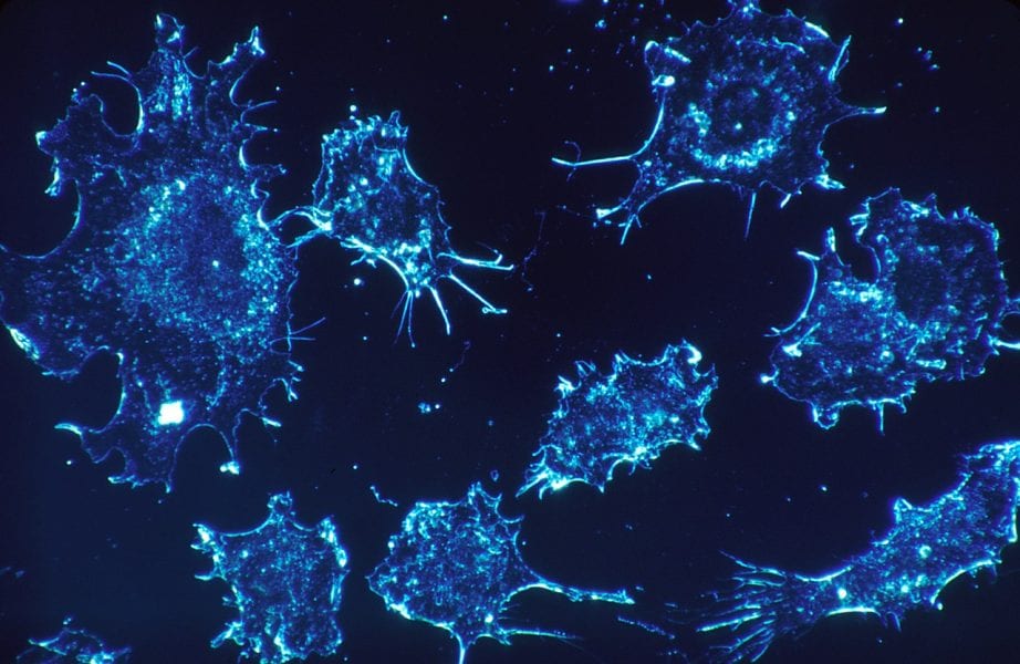
Circulating tumor cells (CTCs) are stray cancer cells that shed from the primary cancer site and travel to healthy tissues and organs via the blood or lymphatic circulation. Once CTCs settle in their new location, they transform healthy cells around them into cancerous cells. Cue cancer metastasis, stage IV cancer. Ever since Dr. S.H. Seal published the first report on the isolation of CTCs in 1964, they have been a quintessential target for biomedical researchers.
Single CTCs have been traditionally considered as the majority of CTCs. CTC clusters were hard to detect in blood flow both in vivo and in vitro. In their recent Editor’s Choice paper in Cytometry Part A, Xunbin Wei and colleagues from the School of Biomedical Engineering of Shanghai Jiao Tong University have demonstrated that the proportion of CTC clusters in circulation increases during cancer metastasis. The goal of this study is to measure CTC clusters in the blood stream of live animals by a two-color in vivo flow cytometry (FCM). Single CTCs and CTC clusters are detected and quantitated in the blood of mice with orthotopic liver cancer and subcutaneous prostate cancer, six times each. The DiD-fluorescently labelled tumor cells are detected confocally in a continuous manner.

Of paramount importance, CTC clusters are easy to detect by in vivo FCM. The dramatic increase of CTC clusters in circulation by 10-40% during cancer progression implies that they are more involved in metastasis than currently perceived. This crucial methodology can be used as a diagnostic measure to estimate the risk of metastasis, and explain the mechanisms of this critical step in cancer progression in a better detail. To learn more about this study’s importance, read this issue’s editorial by Cytometry Part A Editor-in-Chief Attila Tarnok entitled “Measuring the Vicious Ones”.


