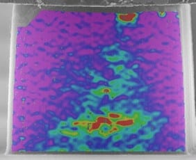
 A thorough understanding of the complex hierarchical mechanics of nanoscale materials systems has been hindered by a lack of experimental techniques capable of measuring deformation of individual nanoscale elements across regions spanning tens to hundreds of microns. Now, researchers at the Air Force Research Laboratory and the University of Michigan have solved this problem using digital image correlation (DIC) to spatially map the deformation and strain of compressed CNT arrays.
The technique utilizes in situ SEM compression and DIC analysis to track the deflection of individual CNTs within defined columns of widely varying aspect ratio. Using it, researchers uncovered a consistent mechanical yield criterion of 5% local strain, although column-level deformation and buckle initiation location varied widely with aspect ratio.
DIC is used extensively for macroscopic materials, but has been limited in application to nanoscale systems due to difficulties in surface preparation of a traceable speckled pattern and availability of compatible in-situ image acquisition techniques. The researchers found that the native topology of CNT forests in SEM micrographs provides a traceable surface speckle pattern for DIC, bypassing the need for surface treatments that may alter their mechanical response. The technique is expected to extend to multiple nano- and micro-scale material systems and enhance model validation and understanding of mechanically coupled responses such as interfacial transport, physical sensing, and energy storage.


