Scientists have now provided real time, label-free images of red blood cells deforming immediately after exposure to high doses of the drug ibuprofen.
“Initially, we were interested in finding the answer to a few simple questions,” explained Peter Nirmalraj, a biophysicist at Empa Laboratories, Switzerland, and a contributor to the study. “Is there a dosage dependent effect of over-the-counter drugs, such as ibuprofen, on red blood cells? If yes, could it be qualitatively and quantitatively capture this effect in a label-free manner?”
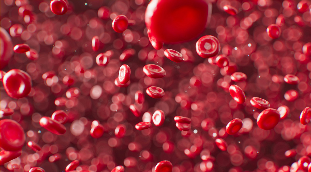
The trillions of red blood cells in our blood have the important job of carrying oxygen to all corners of our body, and play a role in clotting so we do not bleed uncontrollably. Their unique disc or doughnut shape allow them to deform so they can twist and turn their way through the circulatory system.
If red blood cells lose this deformability, blood flow can become sluggish with blockages in small vessels and reduced perfusion to organs and tissues.
Ibuprofen’s affect on blood cells
A well-known disorder affecting red blood cells is anaemia, where a subcategory of this is hemolytic anaemia, a condition where red blood cells are destroyed faster than they can be made. Sufferers are either born with hemolytic anaemia or acquire it through conditions such as auto-immune disorders, burns, or the use of certain medications.
One type of medication that can cause this is ibuprofen, which belongs to a class of drugs called nonsteroidal anti-inflammatory drugs (NSAIDs). While a potent pain killer, ibuprofen has been known to change red blood cell membranes, as demonstrated by one previous study that used snapshot images. Up until now, the inability to take live images to track the process has meant it has been unknown if such changes reversed.
“[If] we can clarify the origin behind such an effect, that would be useful as a complementary indicator for clinicians to identify through blood analysis the effect of drugs on human health,” said Nimalraj.
DHMT provides real-time imaging
To solve this problem, Nimalraj and his team turned to a cutting-edge, commercially available technique called digital holotomography (DHTM) that allows for real time imaging of live cells. This means cells can be visualized in their native state without the need to be labelled with radioactive or fluorescent probes, providing far more detail than before, even visualizing components within cells.
Brought to life by this advanced imaging technique, red blood cells could be seen forming projections called spicules on their membrane when exposed to ibuprofen in a Petri dish. The high ibuprofen concentrations used in the study relate to doses the researchers say should never be taken without medical prescription, whereas low concentrations correspond to doses typically used on a day-to-day basis.
The researchers observed that cells exposed to low concentrations of ibuprofen returned to normal shape within 20 minutes, but no reversal was seen in higher concentrations, even after 1.5 hours.
The high concentrations of ibuprofen caused more red blood cells with spicule projections to form than normal red blood cells. To be sure these spikes were from ibuprofen and not another cause, the team performed control experiments using urea and distilled water and, as expected, no spicule projections formed.
But why and how is this happening? The team believes the answer lies in the negative charge of the ibuprofen particles that interfere with the outer lipid layer of the red blood cells membrane, deforming it. Furthermore, the DHTM imaging reveals the spicules can move and even split into daughter spicules.
These observations were seen in both healthy red blood cells as well as cells affected by sickle cell anaemia, a disorder in which a genetic mutation causes a sickle shaped red blood cell, affecting their ability to bend, reducing blood flow. However, there is no evidence to say this happens in healthy individuals taking the drug.
A gateway to study other blood disorders
It’s important to note that safety profiles for long- and short-term use of ibuprofen are well established, with data going back as far as 1969 when it first entered the market. While the observations made in the current study are interesting and warrant further exploration, the health effects of this phenomenon have not been explored in a real biological setting.
The current study’s focus was instead on demonstrating the utility of DHTM imaging technique to visualize the effects of drugs on cells in the lab in real time, with greater detail than before.
“Our work demonstrates the usefulness of DHTM as an analytical tool for monitoring dosage dependent effects of medicines on blood, which can be integrated with clinical pipeline for assessment of treatment efficiency and response of a patient to prescribed drugs through screening of blood,” said Nirmalraj.
This real time imaging is the gateway to researching a host of blood disorders, including sickle cell disease, thalassemia, and diabetes, to help determine disease stage and treatment effects. More than that, the team say their technique has diagnostic potential to identify diseases that alter red blood cell shape, including malaria and even neurocognitive disorders.
Reference: Damien Thompson, Peter Niraj Nirmalraj, et al., Label-Free Digital Holotomography Reveals Ibuprofen-Induced Morphological Changes to Red Blood Cells, ACS Nanosci. Au (2023). DOI: 10.1021/acsnanoscienceau.3c00004
Feature image credit: ANIRUDH on Unsplash


