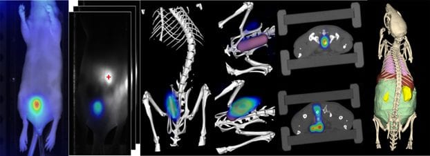
Fluorescence-mediated tomography (FMT) is a promising imaging technology which allows three-dimensional assessment of the fluorescence distribution in deep tissue regions of the body. FMT can be combined with an anatomical modality, a micro-computed imaging tomography such as µCT, which enables more accurate and user-independent analysis because the µCT makes the images easier to interpret particularly for deep locations. Although FMT application is currently limited to rodents or parts of the human body such as hands and breasts, it already proved itself to be a very interesting and promising technique.
Stefanie Rosenhain and other scientists from the Institute for Experimental Molecular Imaging (ExMI) in Aachen, Germany recently performed a study to assess sensitivity and accuracy of µCT-FMT under realistic in vivo conditions in deeply-seated regions. They acquired fluorescence reflectance images (FRI) and µCT-FMT scans of mice which were prepared with rectal insertions with different amounts of fluorescent dye. The improved FMT reconstruction could detect more accurately and sensitively as opposed to FRI and the original FMT reconstruction, plus it had a significantly improved signal localization as well.
With these results it was demonstrated that µCT-FMT is a highly sensitive, non-invasive procedure that provides accurate information in deep-tissue regions and might be a great tool in preclinical imaging studies on colon cancer and inflammatory bowel diseases. The established protocol the team set up, is helpful to assess sensitivity and accuracy for evaluating future improvements of the fluorescence reconstruction. The protocol is simple and reproducible too: due to the complex tissue properties and the presence of motion, an in vivo protocol is important for realistic assessment of sensitivity and accuracy, so the team used commercially available devices and a commercially available probe (OsteoSense) for their experiments to enhance the reproducibility for other research sites. Rosenhain and her team hope that a better understanding of the sensitivity and signal localization will help with planning and conducting future preclinical experiments using this interesting imaging technology.


