Imagine having a camera that could capture and image every molecule inside a cell. We would be able to easily monitor their function and capture any mistakes in their regular activity at any time point.
Detecting such mistakes would be crucial as many lead to defective protein synthesis, defective cell function, and eventually (in some cases) organ failure. While researchers do not possess such powerful cameras, they have developed creative solutions to gain the ability to see inside of cells.
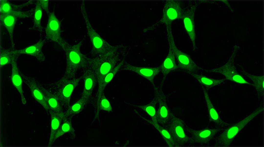
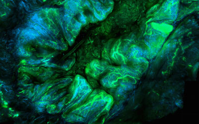
“In reality, we capture images of biomolecules inside of cells using a fluorescence microscope and molecules labelled with a fluorescent dye,” says Professor Nikhil R. Jana, at the School of Materials Science at the Indian Association for the Cultivation of Science (IACS). “Labeling them with the dye makes them visible under a microscope, and microscopy allows magnified imaging at subcellular length scale.”
While this method has allowed us to peak into and study cells, these dyes have limitations. They bleach/react under continuous light exposure — which is a requirement for imaging —and the images fade with time.
In last 25 years, researcher have been developing fluorescent-nanoparticle-based labels that overcome these limitations: they provide bright images and do not bleach under light exposure, so that every intracellular activity can be precisely imaged via fluorescence microscopy.
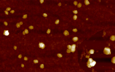
As a result, in recent years chemists have become very excited about fluorescent-carbon-dot-based labels for wider biomedical applications. Compared to large fluorescent tags, fluorescent carbon dots are tiny emissive particles composed of a carbon core that is 1-10 nm in size surrounded with an oxygen rich surface. Their emission can be tuned from the visible to near infrared region just by changing their size — this is difficult for molecular probes as this would require changes in their chemical structural — and are ideal for wider biomedical applications due to the absences of any metals in their composition as metals can be toxic when introduced to the cell or the body in general.
“Using already established chemical reactions, one can transform a synthesized carbon dot into fluorescent label that is useful for imaging specific components of cells including metal ion, acidic/basic environment, proteins, and enzymes,” adds Jana. “Toward this end, extensive research is ongoing in the development of carbon dots with a specific sizes that emit only one color and can image intracellular targets without any interference from background fluorescence. In addition, a carbon dot-based label that selectively images a target biomolecule is highly important.”
Now, in a recent study published in Wires Nanomedicine and Nanobiotechnology, Jana and co-workers propose that fluorescent carbon dots could, in fact, be more useful than we might have originally thought.
“The critical requirements for intracellular imaging probes are that they are small in size (preferably < 10 nm), easily enter the cell, and interact with specific compartments and/or molecules,” he adds. To meet these requirements, fluorescent-carbon-dot-based bioimaging probes have been developed that are 2-10 nm in size with tuneable visible emissions. This means that they can easily be applied like molecular probes but with better performance.
More importantly, they can be synthesized from biomolecules (e.g., carbohydrates, amino acids, and vitamins) via simple carbonization or colloid-chemical approaches and then can be appropriately functionalized for targeting/imaging to different cellular components (e.g., lysosome, mitochondria, nuclei and nucleolus), chemicals (e.g., protons, metal ions, or superoxide species), or biomolecules (e.g., glutathione, amino acids, and DNA).
“Future research should focus on the synthesis of carbon dots with narrow particle size distribution, strong red/near-infrared emissions, and appropriate design and functionalization for subcellular targeting,” the authors stated in their study. This is because many reported carbon dots are currently lager than the desired range of < 10 nm and the majority of reported carbon dots have blue/green emission, but red/near-infrared emission is preferred to minimize background fluorescence of cell/tissue environment. “We expect that in coming decade, more advanced intracellular imaging probes will be developed with the advantages over conventionally used semiconductor quantum dots,” the authors concluded.
Research article found at: Haydar Ali, Santu Ghosh, Nikhil R. Jana, WIREs Nanomedicine and Nanobiotechnology, 2020, doi.org/10.1002/wnan.1617
Kindly contributed by the authors


