Kidney cancer affects 65 000 new patients every year with more than 13 000 deaths. As cross‐sectional imaging has become ubiquitous, the number of small, incidentally detected renal masses has increased. The gold standard treatment of renal masses is extirpative surgery with renal masses biopsy (RMB) reserved for specific cases. This paradigm results in significant overtreatment of renal masses, as is evident by the nearly 6000 benign tumors each year which are misclassified radiographically and removed surgically.
Since overtreatment of renal masses exposes the patients to unnecessary risks – like bleeding, infection and tumor seeding -, improving for the accuracy of preoperative diagnosis could be beneficial.

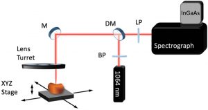
SWIR RS system. BP, band pass filter; DM, dichroic mirror; LP, low pass filter; M, mirror
A team of researchers from the US see a solution in Raman spectroscopy (RS). RS is a mature technique for optical characterization of the compositional properties of materials that has demonstrated promise as an in vivo, label‐free, real‐time, nondestructive diagnostic tool in many malignancies. The team hypothesized that the use of Short wave infrared (SWIR) RS for renal masses diagnosis may overcome limitations related to high background autofluorescence previously observed in RS studies of the kidney.
In their study they demonstrated that the combination of SWIR RS with advanced outlier detection and pattern recognition algorithm accurately differentiated normal and malignant kidney tumors. Doing so, RS has the potential to be used as a diagnostic tool in kidney cancer and for intraoperative guidance during partial nephrectomy. Further larger sample sizes and technical improvements are still needed to unlock this method’s full potential.


