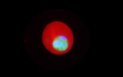
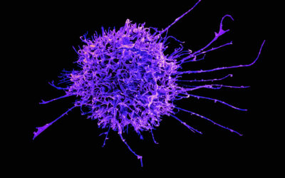
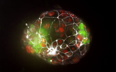
In a continuous effort to understand how genomic DNA structures accommodate specific gene activity and cell function, Szydlowski, Go, and Hu review some of the new super-resolution imaging tools and fluorescent labeling strategies for visualizing the nanoscale chromatin structure in WIREs Systems Biology and Medicine.
Close packing of DNA molecules poses significant challenge for fluorescence microscopy. When visible light is used to visualize nanoscale DNA structures, the diffraction limit of approximately 200 nanometers blends sub-diffraction DNA structures into a blurry spot. This process is analogous to using a ruler, whose smallest unit of measurement is 200 nanometers, to measure the size of objects smaller than 200 nanometers. Even though we can still “measure” the DNA structure, we cannot obtain useful spatial information due to the resolution limit.
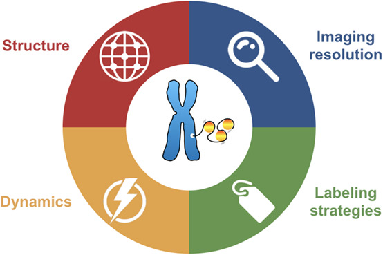
Thanks to the development of super-resolution imaging techniques, we have been able to expand our ability to view the fundamental structures of chromatin in 3D space and time. However, unlike traditional fluorescence images, interpreting super-resolution data requires a careful understanding of the working principles of the imaging modality, labeling quality of the sample, and potential artifacts.
Three separate super-resolution techniques are reviewed by Szydlowski, Go, and Hu, with emphasis on chromatin imaging. In addition to the working principles, they discuss some practical imaging aspects, such as phototoxicity for live imaging and sample fixation artifacts. Fluorescent labeling techniques are also summarized into four major groups, and recent developments in correlated imaging techniques are briefly surveyed, providing a valuable reference to guide experimental design and data interpretation in the field of chromatin imaging.
Kindly contributed by the Authors.


