Embryonic brain development is a complex process, which involves highly regulated cellular dynamics of proliferation, differentiation, and migration. Recent advances in stem cell technology allow researchers to study 3-dimensional human cell cultures, which mimic key aspects of brain development on-a-dish (“brain organoids”). Brain organoids offer the possibility of studying human specific neurodevelopmental disorders, and fundamental questions on the self-organization of the brain during development.
New methods are continuously developed to improve organoid culturing conditions, develop high-throughput capabilities, and allow longer culture periods. However, as the organoid develops, it grows into an opaque millimeter size neuronal tissue, that lacks vasculature. The tissue opacity prevents researchers from performing live imaging studies of organoid development, and the lack of vasculature leads to cell necrosis at the organoid core.
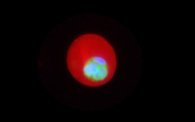
Recently, Karzbrun et al. from the laboratory of Orly Reiner at the Weizmann Institute of Science have developed a micro-fabricated device that supports long-term culture and live imaging of brain organoids. The protocol, published in the latest issue of Current Protocols in Cell biology, offers studying cellular dynamics using live-imaging of in-tact developing organoids, for the first time. The high-resolution imaging can be carried out for weeks without disturbing the organoid growth. The described technique relies mostly on low-cost laboratory instrumentation and reagents, and is compatible with existing organoid culture conditions and commercial microscopy systems.


