Tumor angiogenesis, the formation of new blood vessels that supply the tumor with oxygen and nutrients and aid cancer spread, has been an important target for the treatment of several cancer types. Imaging of tumor vasculature enables the assessment of the efficiency of these antiangiogenic therapies, thereby facilitating their transition from bench to bedside. Numerous imaging modalities have been used for the evaluation of tumor microvasculature properties in response to such treatment by a Dynamic Contrast Enhanced (DCE) approach. However, further efforts are still required for the development of non-invasive imaging tools and contrast agents with less side-effects.
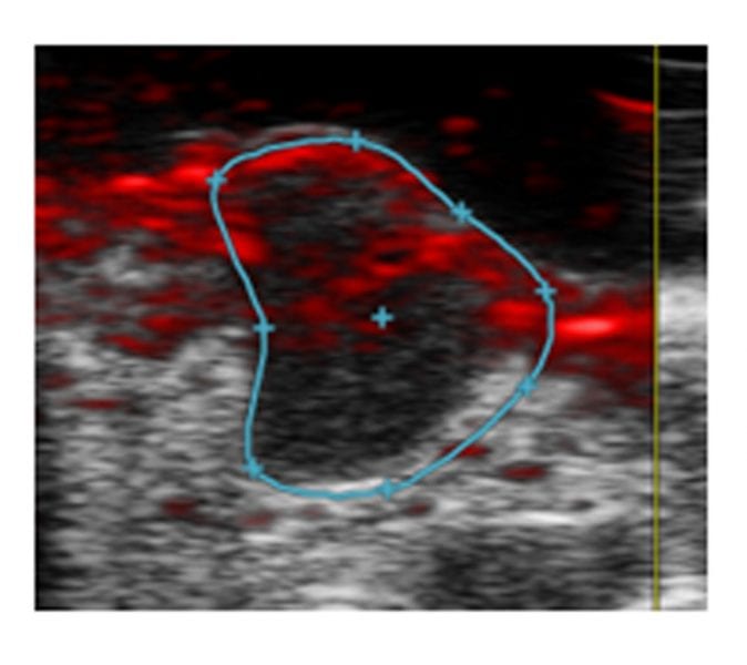
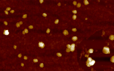
 Researchers from the University of Torino report, in their recent article in Advanced Healthcare Materials, novel systems based on naturally occurring biopolymers as contrast agents for photoacoustic (PA) imaging. These nano-sized, highly water-soluble melanin-based particles are synthesized through a novel simple synthetic strategy. Due to their small size and high water-solubility, they show good biocompatibility with rapid clearance, along with good chemical stability. In mice bearing breast tumors, they accumulate in the tumor and demonstrate impressive contrast enhancement. Furthermore, they are efficient as a probe for the evaluation of changes in tumor vasculature exploiting a DCE-PA approach, as proven following treatment with a drug that increases tumor vascular permeability. The authors foresee that when modified on the surface, these nanoparticles are capable of in vivo active targeting of specific molecular markers.


