The actress Marylin Monroe suffered from endometriosis her whole life and endured multiple surgeries and miscarriages due to the condition. It is suggested that her tragic death from a pain medication overdose was due to chronic endometriosis pain.
Endometriosis is a painful, chronic disorder affecting 10-15% of reproductive age women and girls. It is defined by the abnormal growth of endometrial-like cells outside of the uterine cavity. The heterotopic tissue bleeds each menstrual cycle, which results in the formation of abdominal lesions and extensive adhesions. The growths can damage the uterus and its adjacent organs, including the bladder, bowel, fallopian tubes, and ovaries, resulting in infertility in 30-40% of cases.
The causes of endometriosis are poorly understood. The average time from the onset of symptoms to definitive diagnosis is 10-12 years. Delayed diagnosis is due to a current lack of non-invasive diagnostic methods, and the only absolute way to identify the condition is by laparoscopic surgery.
Current medical therapies prevent fertility (e.g. contraceptive pill or progestin-releasing IUD) and can cause side effects resulting in diminished quality of life. Therefore, many patients seek surgical removal of the lesions. Unfortunately, post-surgical recurrence rates are high, and long-term success largely depends on the skill of surgeons in locating and efficiently removing the abnormal tissue.

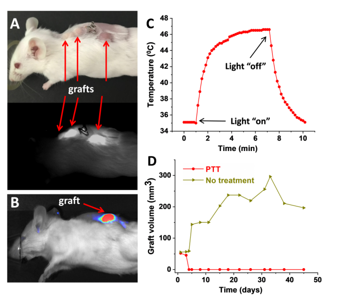
In a recently published article in Small, a group of scientists from the USA led by Oleh Taratula and Ov Slayden describe a novel nanoplatform that identifies endometriosis tissues using real‐time near‐infrared (NIR) fluorescence and ablates them via photothermal therapy (PTT). The nanoplatform comprises polymeric nanoparticles carrying silicon naphthalocyanine (SiNc) which activates the fluorescence signal following internalization in endometrial cells and ablates them by increasing cellular temperature to 53 °C upon interaction with NIR light.
In studies using biopsies of endometrium and endometriosis transplanted into immunodeficient mice, the researchers showed that, 24 h following intravenous injection, the nanoparticles accumulate in endometriotic cells to define the endometriotic grafts with NIR fluorescence. This demonstrated the remarkable diagnostic potential of the developed nanoplatform. Moreover, the nanoparticles increase the temperature of endometriotic grafts up to 47 °C upon exposure to NIR light, completely eradicating them after a single treatment.
These results hold real promise in improving the efficacy of endometriosis ablation therapy and therapeutic potential to reduce the high rates of recurrence after surgery, and potentially in developing non-invasive diagnostic tools for an early detection of Endometriosis.
Reference: Abraham S. Moses, et al. ‘Nanoparticle‐Based Platform for Activatable Fluorescence Imaging and Photothermal Ablation of Endometriosis‘. Small (2020). DOI: 10.1002/smll.201906936


