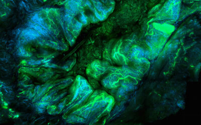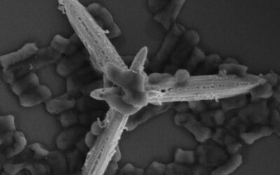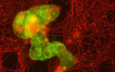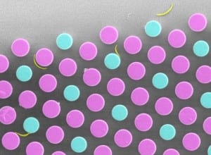
Electron microscopy image showing Plasmodium sporozoites (yellow) between regularly structured micropillars of different sizes (magenta: 10 µm thick; cyan: 8 µm thin).
Malaria parasites are transmitted by mosquito bites. During the bite the parasites are deposited in the skin and immediately start to move at very high speed through the tissue to find and enter a blood vessel. This first step is essential for establishing an infection that can lead to malaria.
The parasite forms deposited by mosquitoes are called Plasmodium sporozoites and are elongated curved single cells. Curiously, these sporozoites move in circles similar to their curvature in vitro. In vivo imaging has revealed that all sporozoites that enter into the bloodstream do so by choosing the small capillaries, but not the large blood vessels that are also present in the skin.
Freddy Frischknecht (Heidelberg University Hospital) and Joachim Spatz (MPI for Medical Research and University of Heidelberg) have revealed that the curvature and circular movement are linked to finding better ways into small capillaries. In their study, published in Advanced Healthcare Materials, they have generated an artificial environment where blood vessels are mimicked by arrays of micro-sized polymer pillars. Adding the parasites to these arrays allowed them to observe their behavior and association with pillars of different sizes and shapes.
Intriguingly, the parasites preferred to associate with structures of a curvature similar to their own, which is at the same time the diameter of small blood vessels within the skin. The study shows not only a general utility of mixed-pillar arrays for the investigation of motile cells with their environment, but also proposes a ‘structural tropism’, which the malaria parasite has developed to help it find and enter blood capillaries.


