Transmission Electron Microscopy (TEM) is a neat tool for the investigation of all kinds of materials, surfaces and biological samples. Over the past decades technological innovations have made this method suitable and affordable for most analytical labs. However, not all sample preparation techniques are suitable for every kind of material. If you have worked with TEM you might know that it is a technique quite prone to cherry-picking. Artifacts can easily be ignored; drying patterns can be misinterpreted as structural features, etc., because it better fits the preconceived scientific narrative. Therefore, for TEM, like every other experimental technique, proper training is necessary. You have to acquire a minimum knowledge of the physical principles involved in order to be able to draw the right conclusions from the samples you are investigating.
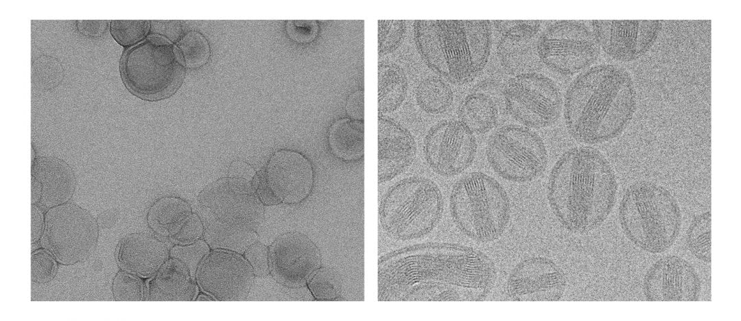
Linda Franken et al. from the University of Groningen in the Netherlands conducted a literature survey and found that in a lot of scientific papers, transmission electron microscopy is carried out in a questionable way when assessing the structure of self-assembled organic objects in solution. In their recent paper in the open access journal Advanced Science the authors give a guideline on how to apply the three most frequently used sample preparation techniques for TEM of soft matter, namely drying, staining, and cryo-TEM.
The case studies show that drying is explicitly not recommended for the vast majority of cases. Nevertheless, analysis of studies using these techniques published within 2010-2015 showed only a moderate uptake of cryo-TEM experiments, which is around 29%. These results underline the authors’ wish to change the perception of drying as a preparation method for soft matter samples and guide researchers towards the more appropriate cryo-TEM technique.

