“Science is a harmonious blend of beautiful imagery and pure reasoning.” (V. S. Ravi) Though science can seem dry and technical at times, there is an understated beauty to it — an empirical way of exploring the universe driven by ingenuity, creativity, and drive to understand the phenomena that move the world around us.
A snapshot of this sentiment is captured in this image gallery, the pictures of which were selected by our editors from among our journals for their striking beauty and the innovative discoveries they showcase. From microneedles to treat the eye to uncovering how cancer cells camouflage to survive, this edition of Science in pictures highlights some intriguing studies captured on “camera”.
Feature image credit: André R. Studart, published in Advanced Materials.
Swaying bacteriophages
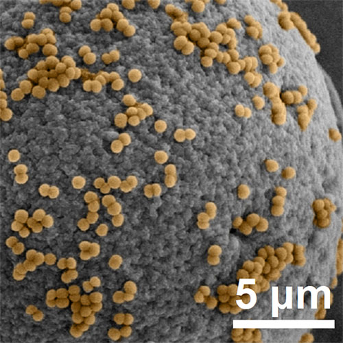
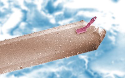
This SEM image shows Staphylococcus aureus bacteria captured on a reactive surface using modified M13 bacteriophages — a type of virus that infect bacterial cells. Publishing in Small by Ting Yang of Northeastern University in China and coworkers, the team describes an approach to create a dynamic and deformable nanointerface based on the unique features of M13 phages in order to create an ultra-sensitive bacteria detector.
The M13 phages can be genetically engineered to help anchor them to the detector’s surface via flexible fibers. This provides an accelerated effect as a result of the swaying of M13 nanofibers, endowing faster kinetics or interactions between the bacteria and the nanosurface to provide a higher sensitivity to the detector.
A bottlebrush chessboard
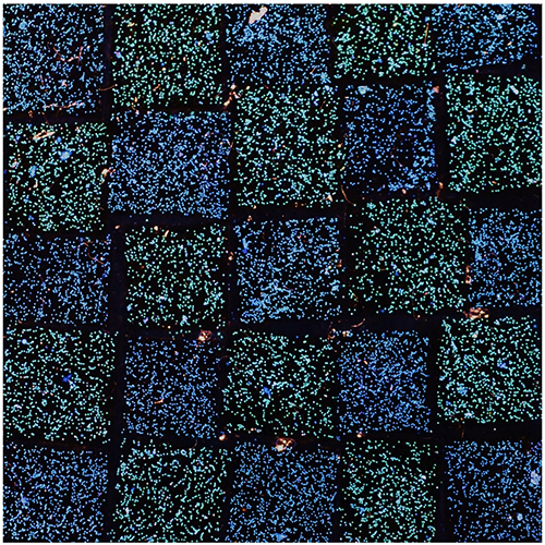
In a study published in Advanced Materials, a team led by Richard Parker and Silvia Vignolini from the University of Cambridge demonstrates how a thermal annealing approach can be applied to photonic pigments made using bottlebrush block copolymers, allowing their colors to be tuned throughout the visible spectrum.
This image depicts polyacrylamide hydrogel containing the different pigments assembled into a checkered pattern. “We demonstrated that thermal annealing can be applied as a discrete step, allowing for thermo-responsive coatings or smart labeling from biocompatible photonic pigments,” wrote the team.
Nanoneedles to treat the cornea
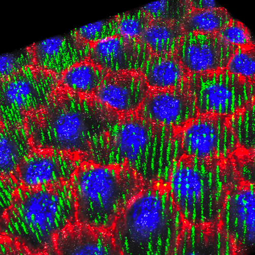
Treating damage to a cornea is difficult, with conventional topical routes being hampered by low drug bioavailability, requiring large dosing, frequent administration, and risks of severe side effects. According to a publication in Advanced Science by Ciro Chiappini of King’s College London, Eleonora Maurizi of the University of Parma, and their co-workers, silicon nanoneedles integrated in tear-soluble contact lenses are an efficient and painless solution for long-term delivery of drugs to the eye.
The 5 µm thickness of the corneal endothelium monolayer matches the length of the nanoneedles, ensuring that other cells in the underlying tissues remain unaffected. This image shows a confocal microscopy image where the nanoneedles localize with cells, indicating successful interfacing.
D4 dice
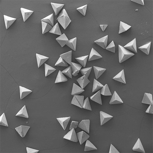
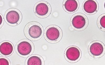
These polyhedral milliparticles are only micrometers wide. They have promising applications in self-assembly, tissue engineering, mechanical engineering, and photonics, where their shape and size have great impact on their function. Creating milliparticles with high symmetry, however, is challenging.
In this study, published in The Journal of Polymer Science, a team led by Shuaishuai Liang from the University of Science and Technology Beijing and Haosheng Chen of Tsinghua University, Beijing, China report a facile means of fabricating polyhedral milliparticles, such as these pyramids, with adjustable shapes and sizes using a microfluidic approach.
Thyroid-on-a-chip
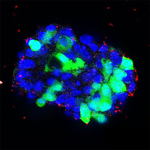
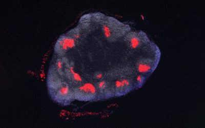
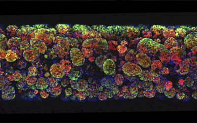
Thyroids are glandular tissues which can be severely affected by endocrine disrupting chemicals. Current in vitro assays to test endocrine disruption by chemical compounds are largely based on 2D thyroid cell cultures, which often fail to precisely evaluate the safety of these compounds.
Publishing in Advanced Healthcare Materials, Stefan Giselbrecht from the MERLN Institute for Technology-Inspired Regenerative Medicine at Maastricht University and collaborators report the development of a thyroid-on-a-chip based on a 3D cell culture “to better recapitulate the thyroid follicular architecture and functions and help to improve the predictive power of such assays”.
Peas and carrots
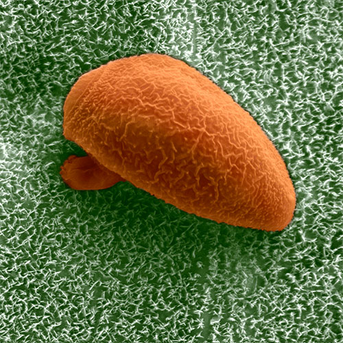
Powdery mildew is a common fungal disease of plants and one of the most important plant pathogens due to its capacity to drastically reduce crop yields. Powdery mildew disease can be caused by hundreds of different species of fungi; although the infections are similar in appearance, the causative fungi are frequently highly specific to their host plants.
A recent study published in Small Science by a team led by Kerstin Koch of Rhine-Waal University of Applied Sciences highlighted the influence of the physical and chemical structure of plant waxes on the successful germination of spores of Blumeria graminis (shown in the micrograph above), a powdery mildew fungus of wheat.
Ultra-miniaturized microfluidics
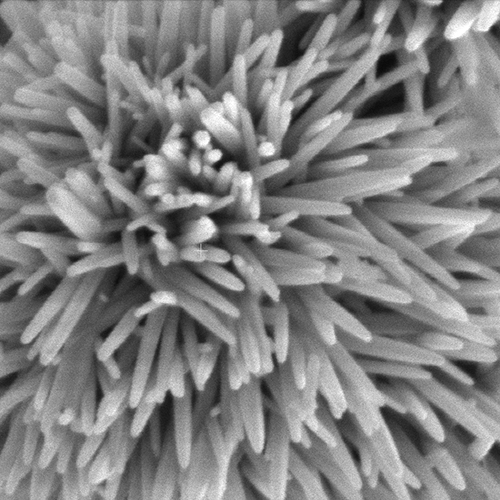
Trace analysis of neurotransmitters in body fluids is critical for early diagnosis of diseases associated with the nervous system, say researchers led by Koji Sugioka from the RIKEN Center for Advanced Photonics in Saitama, Japan. Publishing in Small Science, the team created a portable microfluidic chip, which uses zinc oxide nanowires synthesized on gold film (SEM pictured above) as working electrodes to detect neurotransmitters.
The whole chip was fabricated using “ship-in-a-bottle” technique based on hybrid laser processing that allowed them to directly integrate micro-sized components inside closed microfluidic channels.
Ultralight steel monolith
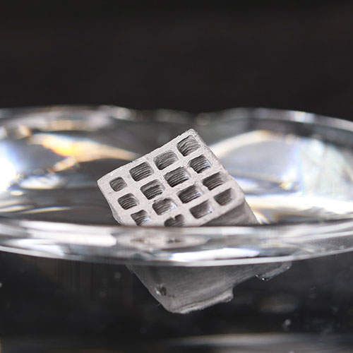
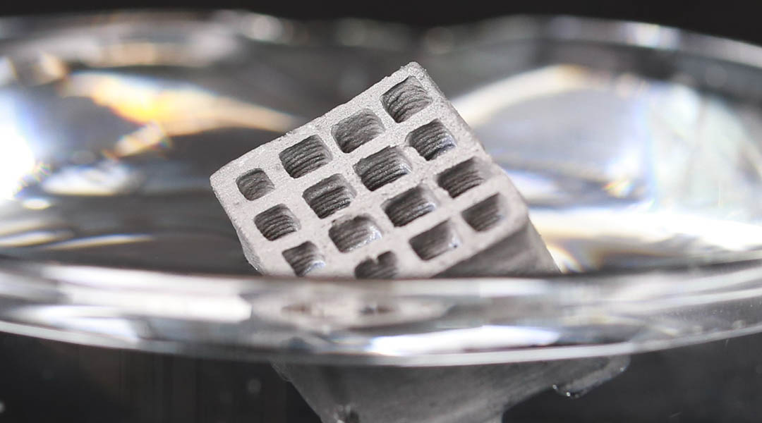
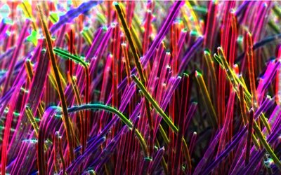
This image depicts hierarchical porous material made from steel. Published in Advanced Materials by André Studart from ETH Zurich and his co-workers, the study describes a means of making centimeter-scale structures like this, which display high mechanical efficiency and reversible, self-reinforcing properties.
Adaptive response, such as self-stiffening under force, is a hallmark of biological materials, making these monoliths highly desirable light-weight functional materials.
Nanofiber networks
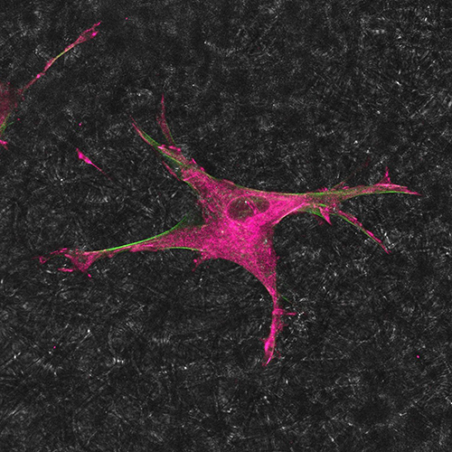
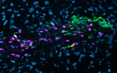
The growth, replication, and differentiation of cells is intimately connected to their external milieu — a complex mixture of proteins and other molecules known as the extracellular matrix (ECM). This image shows the fluorescence signals of actin (green) and paxilin (magenta) components of the intracellular cytoskeleton that play important roles in modulating cells’ reactions to signals received from the ECM.
A team from Sichuan University created a network of curved nanofibers mimicking the ECM structure to demonstrate the importance of these curved structures in the mechanical transduction and subsequent differentiation of stem cells, publishing their results in Advanced Science.
Cancer cells that camouflage to survive
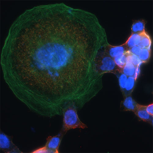
Radiotherapy is vital in cancer treatment, with approximately half of cancer patients receiving radiotherapy at different periods during treatment. Irradiated cancer cells often grow much bigger than normal, as is common for cells that are about to die, and is taken to be a very good sign that radiation had the desired effect.
But a team of researchers led by Chuanyuan Li from Duke University, Yongyong Shi and Qian Huang of Shanghai Jiao Tong University School of Medicine determined that some cancer cells elude researchers by camouflaging as normal cells. Recent findings published in Advanced Science show that these “giant” cells could survive a bout of radiotherapy, and after a long enough wait, they could even repair themselves and become almost normal. This will have significant implications in avoiding relapse after radiotherapy.

