It is widely known that female fertility declines with age, and for women who choose to have children later in life comes an increased risk of complications, such as an inability to conceive, birth defects, and miscarriages. According to experts, this can be primarily attributed to the natural decline of egg cells called oocytes.
“It is known that a decrease in oocyte quality is the major cause of fertility decline in aging women,” said Lucia Lagrutta, a senior embryologist at Grupo IARA Procrearte, a clinic for reproductive medicine in La Plata, Argentina.
Researchers have been interested in understanding the underlying biological processes that lead to this decline, which also affects the success of in vitro fertilization or IVF for short.
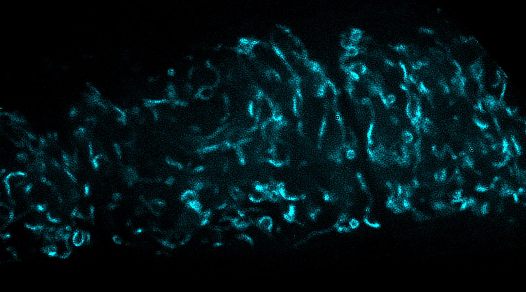
Most notably, some studies have linked the behavior of sub-cellular structures called mitochondria, the power houses of cells, to oocyte quality. “Mitochondrial activity is strongly associated with oocyte quality, including organellar fission and fusion, and changes in their distribution along the oocyte cytosol,” said Lagrutta.
But scientists and physicians are still unsure as to why and how these changes in mitochondria happen as women age.
This is what Meng Wang, senior group leader at Howard Hughes Medical Institute’s Janelia Research Campus in Virginia, United States, and a team of researchers sought to understand in a new study published in Developmental Cell wherethey explored the cellular pathways that become affected in the mitochondria of oocytes.
What can worms teach us?
Finding a good model for studying reproduction in a lab setting is not a simple task, especially when it comes to biological processes that take place over long periods of time.
Wang and the team opted to work with a type of worm known as Caenorhabditis elegans (C. elegans). “While mice have an average reproductive lifespan of eight to 12 months, C. elegans reproduce for only three to six days,” said Wang. “This rapid reproductive cycle allows researchers to quickly examine the impact of various interventions on reproductive aging and gain a deep understanding of how they work.”
While C. elegans are much simpler than humans in terms of their biology, many fundamental biological processes are conserved between the two species, such as cell division, fat metabolism, and even the genetic regulation involved in reproduction.
Additionally, C. elegans and humans have many genes in common, particularly those involved in fundamental cellular processes and pathways, such as those used by the mitochondria to generate energy. Mitochondria take in nutrients and oxygen, and, through a complex chemical process, convert them into energetic molecules called ATP and GTP, which cells use to power various functions.
Taking advantage of this simpler model, Wang and her team studied mitochondria metabolism in aging C. elegans’ oocytes. They found that the production of GTP was increased in older oocytes, and when it was artificially decreased, the scientists were able to prolong the reproductive lifespan of the worms.
This came down to an enzyme called mitochondrial GTP-specific succinyl-CoA synthetase (SCS), which is involved specifically in GTP production and not ATP. When the scientists inhibited this enzyme, the percentage of seven-day-old worms that reproduced was over 70%, while normally at this stage in their life cycle, this would be less than 50%.
“We have established a link between mitochondrial GTP metabolism and reproductive aging for the first time,” said Wang. “When the mitochondrial GTP level is reduced in the reproductive system, animals can maintain their reproductive abilities for a longer period.”
Location also matters
Although mitochondrial activity affects oocyte quality, many studies reported that where the mitochondria exist within the cell’s cytoplasm is also important, showing that when mitochondria accumulate around the cellular nucleus, the oocyte quality is affected. Currently, clinicians and researchers assess mitochondrial cytoplasmatic distribution in the lab as one of the parameters that determine their quality.
“The age-associated increase in mitochondrial clustering near the nucleus is a conserved phenomenon in oocytes across different species, and that mitochondrial positioning could serve as a good indicator of oocyte quality,” said Yi-Tang Lee, the study’s first author and a graduate student in Wang’s lab at the time of the study and now manager of the Human Stem Cell Core at the University of Wisconsin-Madison.
The scientists found that by decreasing the GTP-specific SCS’s enzyme activity, mitochondria were no longer located around the cell nucleus, and were more dispersed in the cellular cytosol in a similar pattern normally found in healthy oocytes. “We have discovered that mitochondrial positioning in the oocyte is central to reproductive health during aging,” said Lee.
From worms to humans
While this study was conducted on C. elegans, its implications could extend beyond the microscopic world of nematode worms. Understanding the intricate relationship between mitochondrial GTP metabolism with reproductive aging could have profound implications for how we approach reproductive health.
“In the clinic, oocyte quality is often assessed qualitatively and subjectively, relying on human expertise that requires years of training,” said Lee. “In the future, non-invasive imaging of mitochondrial position and subsequent analysis using a computational method similar to the one demonstrated in our work may provide a quantitative and objective means of assessing oocyte quality in clinical practice.”
“Taking into account that in IVF, laboratory fertilization rate and embryo quality decrease significantly in older women, [with the findings of the current study] it seems to be possible to improve IVF outcomes,” said Lagrutta, who was not involved in the study.
“One possibility could be to apply strategies to reduce GTP levels and change mitochondrial distribution on mature oocytes retrieved from aged women by ovum pick-up (OPU) and follicular aspiration,” she continued. “Then, these modified oocytes could be inseminated by ICSI [Intracytoplasmic sperm injection], in the IVF laboratory, reducing fertilization failures and improving embryo number and quality in this group of women.”
Future studies will determine if these results are really extrapolated to humans, but for the moment, this study serves as an important first step. “This new perspective could be a window into future research on women’s fertility preservation for those women who plan to delay pregnancy,” said Lagrutta.
Reference: Yi-Tang Lee, et. al., Mitochondrial GTP metabolism controls reproductive aging in C. elegans, Developmental Cell (2023). DOI: 10.1016/j.devcel.2023.08.019


