Cytometry Part A has recently published a Special Issue on Quantitative Phase Imaging for Label-Free Cytometry guest edited by Elena Holden and Gabriel Popescu.
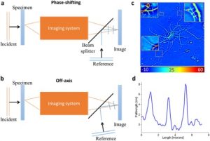
Principles of quantitative phase imaging (QPI). (a) Phase shifting QPI. (b) Off-axis QPI. Both geometries can be implemented in common path and with broad band illumination. (c) Quantitative phase image of a neuron. The insets zoom into the respective selected areas. Color bar indicates optical pathlength in nanometers. (d) Pathlength profile along the line shown in 1c top right inset.
Fixed cell imaging provides a glimpse at a definite moment in the cell’s existence. But one moment doesn’t represent the dynamic processes underlying its function. The biological complexity of a live cell can only be understood when it is placed in the focus of the microscope’s objective. Label-free imaging avoids the limitations inherent to fluorescent probes (phototoxicity, photobleaching) and maintains an appropriate environment for normal cellular behavior.
Quantitative Phase Imaging is a label-free imaging technique that generates optical path difference maps associated with the target specimen. The approach is sensitive to both local thickness and the refractive index of the sample. It is growing in popularity as a complementary technique to traditional cytometry techniques and indispensable to analyze cell dynamics in an unaltered, native state.
The multiple variants of QPI systems can be categorized based on parameters that describe their performance:
- Temporal sampling – acquisition rate.
- Spatial sampling – transverse resolution.
- Temporal stability – temporal phase sensitivity.
- Spatial uniformity – spatial phase sensitivity.
This Special Issue includes the Editorial by Elena Holden, Attila Tárnok and Gabriel Popescu and 13 articles that report on the latest technical developments in QPI used to study the mechanisms of cancer and neurodegenerative disorders, to develop multispecific pharmaceutical formulations, and as a robust segmentation technology for microbial cells.
The issue coincides with CYTO 2017, the 32nd Congress of the International Society for Advancement of Cytometry that will take place at the Hynes Convention Center in Boston, MA from June 10 to 14.

