Bone and cartilage injuries can cause chronic pain, joint stiffness, and impair mobility. Bone and cartilage injuries can result from degenerative arthritis, trauma, or tumor surgery, and often do not heal without major surgery.
Stem cell transplants can directly repair bone and cartilage defects. Stem cell transplants can also reduce pain by producing anti-inflammatory mediators. However, harvesting stem cells from a patient is expensive and requires a biopsy or surgery to harvest them.
Creating commercially available stock supplies of therapeutic cells has many advantages such as instant and abundant availability, consistent quality control and no need of a biopsy to harvest them. Therefore, commercially available, “off the shelf” stem cell products have been increasingly introduced for the treatment of arthritis and other muskuloskeletal disorders.
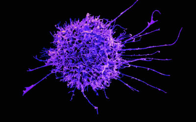
However, “off the shelf” therapeutic cells can be recognized as foreign by the patient’s immune system and lead to their rejection. If an immune rejection is identified within the first week after a cell transplant, drugs that suppress the bodies immune response could be applied to prevent graft failure and support engraftment.
In WIREs Nanomedicine & Nanobiotechnology review, Heike Daldrup‐Link and co-workers discuss how new imaging technologies can help to diagnose an immune rejection of therapeutic cell transplants. These new and immediately clinically available imaging tests rely on using the FDA approved iron supplement ferumoxytol “off label” as a marker for an immune rejection.
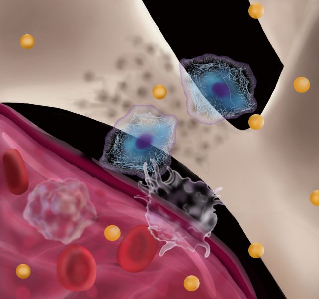
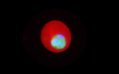
Ferumoxytol is composed of iron nanoparticles, which can be detected with magnetic resonance imaging (MRI). Ferumoxytol can be introduced into either stem cells or immune cells such that the cells can be tracked in vivo with MRI. Labeling the stem cells with ferumoxytol can help to visualize their location and engraftment over time. Labeling immune cells with ferumoxytol can help to diagnose an influx of immune cells into a cell transplant. Both processes can be combined by creating new, second generation cell markers that are linked to enzyme-activatable fluorescent contrast agents. For example, ferumoxytol can be linked to a caspase-activatable fluorescent probe that lights up when therapeutic cells die.
These new imaging techniques can help scientists to better understand stem cell rejection processes and use that information to develop better cell therapies. Since the FDA-approved iron supplement ferumoxytol is immediately clinically applicable through an “off label” use, these imaging techniques can be immediately applied to diagose a failure of therapeutic cell transplants in patients. Since ferumoxytol-MR imaging techniques provide an earlier diagnosis of immune rejection processes than currently available with standard imaging tests, ferumoxytol-MRI might be able to diagnose complications of therapeutic cell transplants at a reversible stage, when medical interventions can still save the transplant.
Kindly contributed by the authors.

