Increasing the resolution of microscopes, by breaking the diffraction limit, can be categorized into near‐field and far‐field operations. The near‐field approaches, such as superlenses, hyperlenses, microsphere-assisted imaging, and near‐field scanning optical microscopy, use evanescent waves containing high spatial frequencies to resolve small objects, but the imaging process needs to be performed very close to the sample. Far-field options, such as emission depletion microscopy (STED), photo‐activated localization microscopy (PALM), and stochastic optical reconstruction microscopy (STORM) are able to provide the resolution at tens of nanometers by selectively activating or deactivating fluorophores, but the pre‐processing and labeling of samples with specific dyes restrict these techniques to biological specimens. Recently, a non‐invasive universal imaging technique, called super‐oscillatory lens (SOL) optical microscopy, was introduced, but this also faces challenges such as a small field of view, the need for complicated nano‐lithographic fabrication of sub‐wavelength features, and the short working distance.
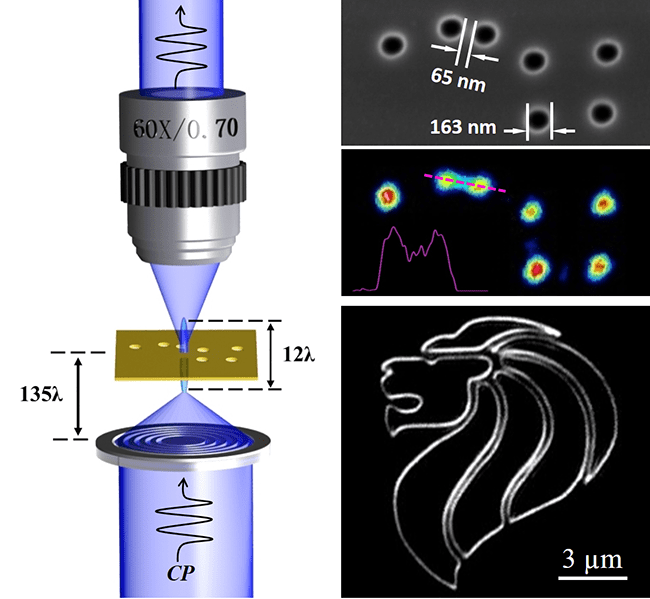
Now, Minghui Hong and colleagues from the National University of Singapore and the Agency for Science Technology and Research (A*STAR), Singapore, report super-critical-lens (SCL) optical label‐free microscopy (see Figure), which clearly distinguishes a pattern with a feature size of 65 nm in air and with a 55 μm working distance. The imaging process is purely physical and captured in real time, does not require any pre‐processing of the samples or mathematical post‐processing of the imaging results. SCL microscopy is able to map the horizontal details of a 3D object through one-time scanning, something that is impossible with other planar lenses and it challenges the received wisdom that the sub‐wavelength imaging requires sub‐wavelength features of the lens. The researchers believe that the method could open the way to wide availability of high‐performance and cost‐effective nanoimaging and nano‐fabrication technologies.

a) When the SCL is illuminated by a 405 nm circular polarized (CP) beam, a sub‐wavelength optical needle 12λ long is formed 135λ away from the SCL plane. The imaging sample is placed in the needle range and raster scanned in the X‐Y plane and the signal is collected by an objective. b) SEM of a nanoscale “Big Dipper” used for the resolution demonstration. c) Imaging result with a normal transmission‐mode (T‐mode) microscope with an NA = 0.9 objective lens under illumination at 405 nm. d) Imaging from laser scanning confocal microscopy (LSCM) with the same wavelength and objective lens. For comparison, the LSCM image is presented with inverted color from the source image, since the LSCM is working in reflection mode which differs from others. e) Imaging result with SCL microscopy which shows that the 65 nm space between the stars ε and ζ can be clearly distinguished. These images are presented by pseudo‐color for clarity. Insets are the line intensity profiles through the center of stars ε and ζ for each image. Scale bars: 500 nm.


