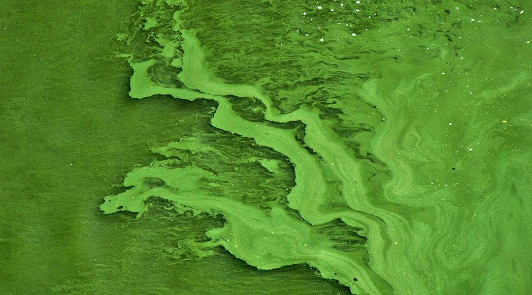
Researchers have long wondered what factors influence algal blooms, which form every year in oceans around the world.
While giant viruses have been known to infect unicellular organisms, including algae, their presumed role in killing off the blooms each year has been challenged by a study published in Science Advances, which has revealed that the relationship between these viruses and their hosts is far more complicated and important than previously assumed.
For Assaf Vardi, a marine biologist, and his team at the Weizmann Institute of Science in Israel, the answer lies in studying single cells rather than the bloom as a whole. “We are the first in the world to be able to track a single cell in natural populations,” said Vardi.
To get answers, the team travelled to Norway to isolate algae from small blooms found in fjord waters near the town of Bergen. There, the University of Bergen has set up marine mesocosms, which are enclosed little ecosystems sealed in submerged bags but separated from the environment.
This provides as “close to natural conditions” as possible while allowing the scientists to control certain variables, such as the water’s salt concentration, and observe the effect on algal growth. “It was a beautiful adventure to the sea, very interdisciplinary, with many people from different countries that were invited to conduct their experiments,” recalled Vardi.
From the lab to the fjords
To study cells on an individual level, Vardi and his team employed a technique called single-cell transcriptomics, which is “very common in the biomedical world but was never tested on natural populations of [algae]” — and took it directly to the sea.
Single-cell transcriptomics measures newly produced messenger RNA, the precursor molecule for the production of proteins, and decodes its function within the cell. By analyzing whether viral RNA molecules were present and which algal RNA was predominantly being produced, the researchers could deduce the state of infection of individual cells.
“This tells us how each individual cell behaves and what makes a cell different in response to an infection from another cell,” said Vardi.
Applying the technique to the fjord mesocosms offered distinct advantages compared to conventional analyses of algae, which are usually performed in sterile labs and on large populations of cells.
The researchers collected and compared several thousand cells from seven little ecosystems and used the data to analyze how healthy the cells were, if they were infected by the giant viruses, and if so, how far the infection had already progressed. The rational being that the effect of an infection on an entire bloom can only be understood by linking scales and analyzing the impact of single, infected cells on blooms comprising millions of cells.
Giant viruses aren’t necessarily the bad guys
Vardi says that he and his team were surprised to discover that the viruses were infecting and killing algae cells on a daily basis — an idea that contradicts previous assumptions that the virus infections were more or less synchronized, bringing an end to the algal blooms all at once every year.
But the daily turnover of algae apparently provides an important function as at the end of infection, the algae cells burst, releasing cellular material into the surrounding water that can be used as fuel by other microorganisms in the ecosystem, providing a continuous flow of nutrients throughout the bloom.
“What we show is that [the release of nutrients] doesn’t happen just at the end of the bloom, but there is a daily turnover of life and death, these viruses infect and kill, and infect and kill,” explained Vardi.
This appears to be quite important for the overall health of the bloom, as the algae depend on bacteria that also flourish in the nutrient-rich environment. Bacteria break down materials the algae otherwise cannot digest, similar to how we depend on the microbiome in our own digestive tract.
Matthias Fischer, a scientist at the Max-Planck Institute for Medical Research in Germany whose work focuses on discovering new viruses in the environment, is excited to see virus-infected cells studied in such detail, on a single cell level.
“If you analyze with bulk methods how an infection progresses, you see the typical profiles from early to progressively late infection states, before the bloom eventually disappears. What [Vardi and his team] see now is that there is a constant underlying infection.”
Fischer, who was not involved in the study, explained that viruses and cells have subtle differences that are easily missed. “You can only appreciate this if you no longer analyze in bulk but go to the single cell level,” he said.
“People like to divide into good guys and bad guys,” added Vardi. “But it doesn’t work like this.”
Expanding to the oceans
“The study provides insights into how we can tease populations apart to see what individuals are doing,” said Steven Wilhelm, professor of microbiology at the University of Tennessee who was not involved in the study. Wilhelm has been studying the ecology of viruses, bacteria, and algae in marine and freshwater samples for over twenty years.
“It remains early days in terms of single cell work,” he said. “Even though one can look at thousands of cells (12,000 single algae in the current case), that is in reality just the algae in about a milliliter of water.
“While amazingly powerful by our current standards, it is likely that improvement in techniques going forward will expand our abilities from thousands to millions.”
By sharing their know-how with the scientific community, Vardi hopes to inspire others to use this technique to eventually study millions of single algae during massive blooms in the open ocean.
This will be vital to understanding the processes under which blooms thrive and then dissipate, which is changing rapidly under the accelerating global climate crisis.
Reference: Assaf Vardi, et al., Daily turnover of active giant virus infection during algal blooms revealed by single-cell transcriptomics, Science Advances (2023). DOI: 10.1126/sciadv.adf7971

