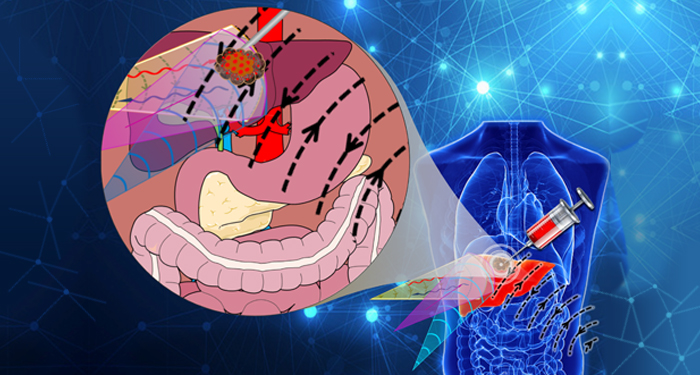
Precise targeting and controlled drug release can significantly enhance the therapeutic efficacy of cancer treatment. Trackable smart drug delivery systems have gained attention in the past decade because they can be visualized by one or more imaging technologies through their distinct physical properties. However, it is still difficult to achieve precise drug delivery because such systems usually rely on a single imaging system that cannot clearly distinguish the drug carrier system from the surrounding tissue and the targeted lesion.
Recently, Huang et al. from the laboratory of Jianhua Zhou at Sun Yat‐sen University, Guangzhou, China, proposed a novel strategy for multimodal visualization of eccentric magnetic microcapsules (EMMs) designed for potential treatment of hepatocellular carcinoma. The EMMs were composed of Fe3O4 nanoparticles and polydimethylsiloxane (PDMS) that could be visualized by magnetic resonance imaging (MRI), computed tomography (CT), and ultrasound (US). Phantom model simulating the in vivo microinjection environment was used to demonstrate that as opposed to using a single imaging system, the integration of imaging data from MRI, CT, and US allowed for precise tracking and positioning of EMMs to the lesion site. In addition, controlled release of doxorubicin from the EMMs could be achieved by applying an external electromagnetic field.
The results, published as the cover article in the latest issue of Cancer Reports, provide a proof of concept that these EMMs serve both as contrast agents that can be visualized by multimodality imaging techniques and as smart drug delivery systems.
To find out more please read: W. Huang, Y. Chen, L. Chen, J. Zhong,A. M. Johri, J. Zhou, Multimodality imaging‐guided local injection of eccentric magnetic microcapsules with electromagnetically controlled drug release. Cancer Rep. 2018, 2, e1154.


